
Clinical Studies and Research
In the lab, a shin reading outperformed a scan of the hip.
A 2026 study in Clinical Orthopaedics and Related Research measured BoneScore in 67 female donors. Each donor was also given a clinical DXA. Then the femur, radius, and L4 vertebra were loaded in a testing frame until they fractured.1
The hip scan was looking directly at the bone that would later break. It could not separate the donors whose femurs held from those whose femurs failed (AUC 0.62; p = 0.08). BoneScore, measured at the shin, did (AUC 0.75; p < 0.001).
Added to a hip density scan, it more than doubled the explained variation in hip strength (28%→52%). In that laboratory testing, BoneScore identified fracture-susceptible femurs better than the hip DXA did (AUC 0.75 vs 0.62).
200+ papers · 65 clinical studies · 70+ institutions
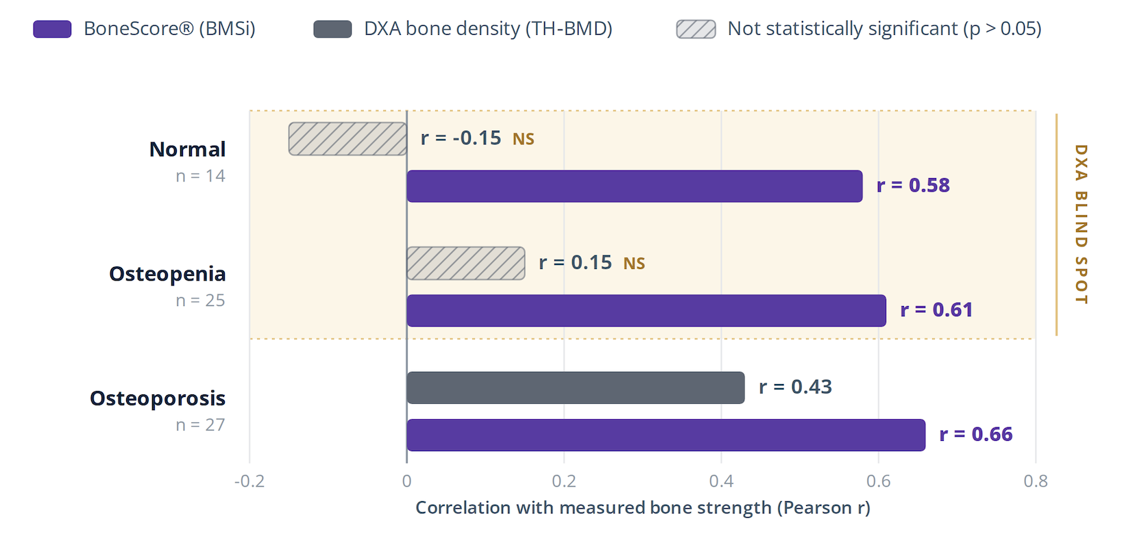
How to read this table: Each bar is the Pearson correlation (r) between a test and measured femur strength, within a DXA-defined group. Higher = the test better reflects true strength. Hatched bars are not statistically significant — the density reading carried no reliable information about strength in that group. BoneScore® was significant in all three groups (p ≤ 0.02).
The bottom line:
- DXA works well at understanding strength when bone density is very low.
- In this cadaver study, BoneScore stayed associated with strength across all subjects regardless of density.
References
- Vaidya RS, et al. Impact microindentation evaluates bone strength, bone quality, and fracture susceptibility across skeletal sites: a cadaver study. Clinical Orthopaedics and Related Research. 2026 (accepted). View source →
